Meet our doctors and staff members
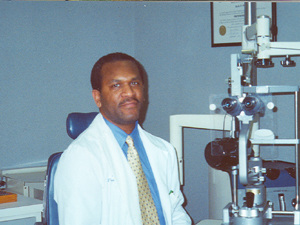
Keith F. Fishe, O.D.
|
Dr. Fishe hails from Bronx, New York and attended Clark College in Atlanta.
A 1987 graduate of the Pennsylvania College of Optometry, Dr. Fishe practices full scope optometry with an emphasis in anterior segment. When not in the office, Dr. Fishe enjoys traveling, reading and golf. |
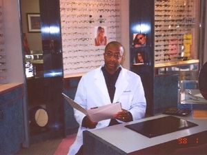
Curtiss Anderson, O.D.
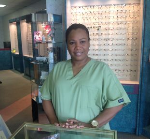
Dianne Cummings- Front Desk Receptionist
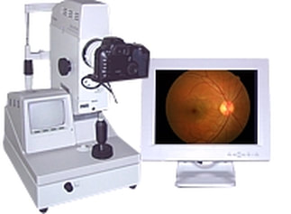
6.5 Digital Retinal Imaging System
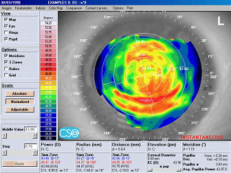
Wavefront Corneal Topography
|
Corneal topography is a computer assisted diagnostic tool that creates a three-dimensional map of the surface curvature of the cornea. An eye with normal vision has an evenly rounded cornea, but if the cornea is too flat, too steep, or unevenly curved, less than perfect vision results.
The greatest advantage of corneal topography is its ability to detect irregular conditions invisible to most conventional testing. |
Corneal topography produces a detailed, visual description of the shape and power of the cornea. This type of analysis provides Drs.Fishe and Anderson with very fine details regarding the condition of the corneal surface. These details are used to diagnose, monitor, and treat various eye conditions. They are also used in fitting contact lenses and for planning surgery, including laser vision correction. For laser vision correction the corneal topography map is used in conjunction with other tests to determine exactly how much corneal tissue will be removed to correct vision.
Computerized corneal topography can be beneficial in the evaluation of certain diseases and injuries of the cornea including:
Corneal diseases,
Corneal abrasions,
Corneal deformities,
Irregular astigmatism following corneal transplants,
Postoperative cataract extraction with acquired astigmatism.
Computerized corneal topography can be beneficial in the evaluation of certain diseases and injuries of the cornea including:
Corneal diseases,
Corneal abrasions,
Corneal deformities,
Irregular astigmatism following corneal transplants,
Postoperative cataract extraction with acquired astigmatism.
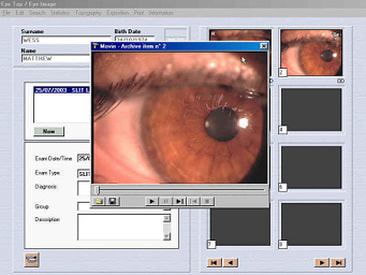
2.0 Slit Lamp Digital Imaging System
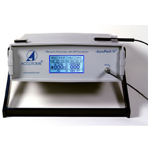
Corneal Ultrasonic Pachymetry
|
A pachymeter is a device that uses ultrasound to determine the thickness of the cornea.
Pachymetry is an essential measurement prior to certain refractive surgical procedures, such as LASIK. Procedures such as LASIK remove tissue from the cornea thus it is important to be certain that the cornea will retain enough central tissue thickness. In addition to laser vision correction, pachymetry is useful in determining the risk of developing glaucoma and interpreting unexpected intraocular pressure (IOP) measurement results. Increased corneal thickness can produce falsely high IOP readings, and decreased corneal thickness can produce falsely low IOP readings. Glaucoma of all types is the second most common cause of legal blindness in the United States and the leading cause of legal blindness among African Americans. Glaucoma usually has no early symptoms, and by the time people experience problems with their vision, they usually have lost a significant amount of their sight. Other uses include: Persons with glaucoma or elevated intraocular pressure, Corneal refractive surgery (pre- and post-operative evaluation), Corneal edema, Fuch's endothelial dystrophy. |